
Purpose. We performed a first-in-human clinical trial. The aim of this study was to determine safety and feasibility of PET imaging with 18F-PARPi in patients with head and neck cancer. Patients and Methods. Eleven patients with newly diagnosed or recurrent oral and oropharyngeal cancer were injected with 18F-PARPi (331 ± 42 MBq) and dynamic PET/CT imaging was performed between 0 min and 25 min post-injection. Static PET/CT scans were obtained at 30 min, 60 min and 120 min post injection. Blood samples for tracer concentration and metabolite analysis were collected. Blood pressure, ECG, oxygen levels, clinical chemistry and CBC were obtained before and after tracer administration.
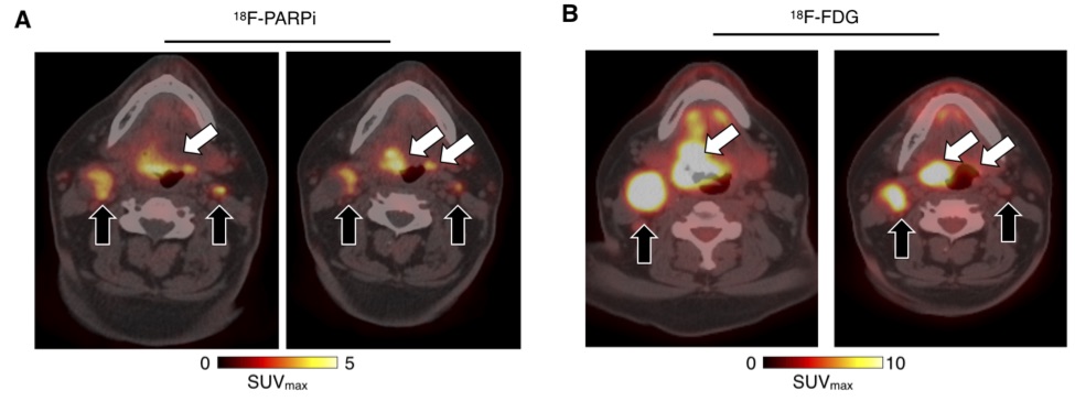
Results. 18F-PARPi was well-tolerated by all patients without any safety concerns. Of the 11 patients included in the analysis, 18F-PARPi had focal uptake in all primary lesions (n = 10, SUVmax = 2.8 ± 1.2) and all 18F-FDG positive lymph nodes (n = 34). 18F-PARPi uptake was seen in 18F-FDG negative lymph nodes of three patients (n = 6). Focal uptake of tracer in primary and metastatic lesions was corroborated by CT alone or in combination with 18F-FDG. The overall equivalent dose with 18F-PARPi PET was 3.9 mSv – 5.2 mSv, contrast was high (SUVmax(lesion)/SUVmax(trapezius muscle) = 4.5) and less variable than 18F-FDG when compared to the genioglossus muscle (1.3 versus 6.0, p = 0.001).
Conclusions. Imaging of head and neck cancer with 18F-PARPi is feasible and safe. 18F-PARPi detects primary and metastatic lesions, and retention in tumors is longer than in healthy tissues.
