
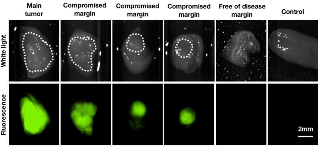
Complete removal and negative margins are the goal of any surgical resection of primary oral cavity carcinoma. Current approaches to determine tumor boundaries rely heavily on surgeons’ expertise, and final histopathological reports are usually only available days after surgery, precluding contemporaneous re-assessment of positive margins. Intraoperative optical imaging could address this unmet clinical need. Using mouse models of oral cavity carcinoma, we demonstrated that PARPi-FL, a fluorescent PARP inhibitor targeting the enzyme PARP1/2, can delineate oral cancer and accurately identify positive margins, both macroscopically and at cellular resolution. PARPi-FL also allowed identification of compromised margins based on fluorescence hotspots, which were not seen in margin-negative resections and control tongues. PARPi-FL was further able to differentiate tumor from low-grade dysplasia. Intravenous injection of PARPi-FL has significant potential for clinical translation and could aid surgeons in assessing oral cancer margins in vivo.
